SQUAMOUS CELL CARCINOMA OF FOREHEAD WITH PERINEURAL INVASION
HISTORY
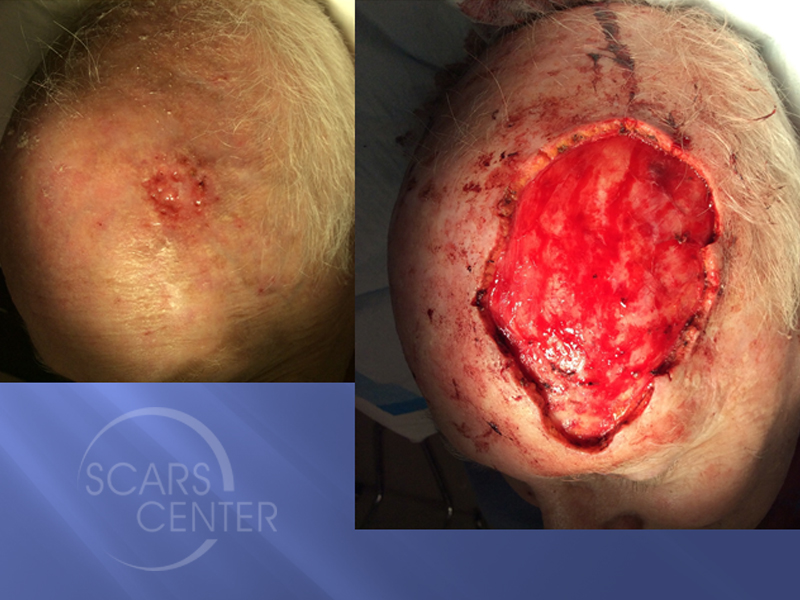
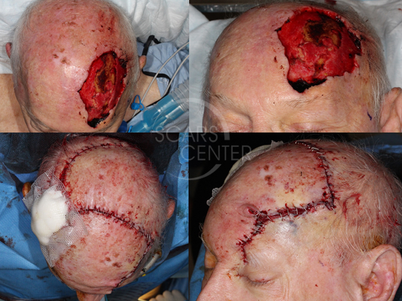
This 81 year old man presented with invasive squamous cell carcinoma with perineural invasion of the left forehead and scalp. Mohs was performed on 2/21/2017. Additional wide margin resection was performed on 2/27/17. It was negative for residual squamous cell carcinoma.
DISCUSSION
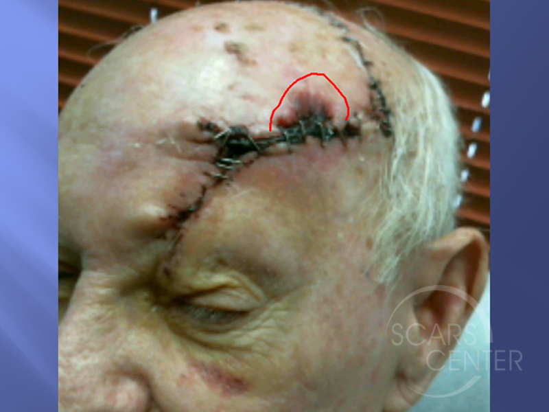
This 81-year-old man presented with invasive squamous cell carcinoma with perineural invasion starting in the left forehead and scalp area. Over seven levels of Mohs excision were performed, tracking out the carcinoma to the supraorbital nerve creating a large defect in the forehead and scalp. Despite negative Mohs margins, it was recommended that additional margins were taken around the supraorbital nerve. This was done due to a simple observation that perineural invasion has been shown to track up to 2 cm from obvious gross tumor involvement. As a result, additional 1.5 cm margins were taken below the lowest extent of the forehead Mohs defect. This involved taking some of the superior orbital portion of the supraorbital nerve to achieve the 1.5 cm margin. These 1.5 cms together with a 0.5 cm margin of the Mohs excision achieved the minimal necessary 2 cm margin clearance of perineural invasion. Fortunately for this patient, no carcinoma was found in the additional supraorbital nerve resection. A large defect was reconstructed with very large rotation flaps and a split thickness skin graft to the donor site of the right opposite scalp. Clearly despite the negative clinical margins, the patient is at high risk of recurrence due to the nature of perineural invasion. Recommendation was made for radiation therapy to the forehead and the scalp, sparing the orbit. Additional 2 to 3 cm margin was recommended around the area of surgical treatment. Finally, serial MRIs of the brain and orbit should be performed every four to six months for the next two years to look for possible recurrence.

