Call Us Today:
949.200.1600

Basal Cell Carcinoma

Home > Diagnosis > Basal Cell Carcinoma

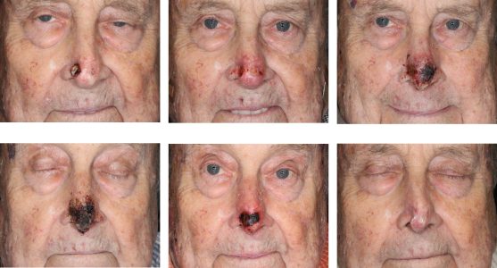
About Basal Cell Carcinoma

Basal cell carcinoma (BCC) is the most common form of skin cancer arising from basal cells residing within the dermal-epidermal junction of the skin. This cancer is usually easily detectable as it forms plaques, nodules, or scabs on the surface of the skin. Basal Cell Carcinomas of the skin are the least aggressive skin cancers that we treat in Orange County. These cancers only cause local tissue invasion and some destruction, and almost never metastasize. Basal Cell Carcinoma of the skin are very slow growing as a rule with some lesions taking many months to evolve.


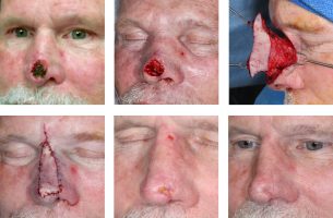
We perform basal cell carcinoma treatment from our Newport Beach clinic. Treatment of these cancers are numerous including curettage, simple excision, Moh’s surgery, and superficial radiotherapy. Other alternative treatments can include topical cancer treatment creams.

Diagnosis

A lesion presenting as a non-healing skin lesion is biopsied. BCC can be diagnosed with a simple shave biopsy of the skin lesion. Punch and incisional biopsies can also be done. Diagnostic non-invasive techniques include Reflectance Confocal Microscopy (RCM). Most commonly used biopsy technique is a shave biopsy. Even very thin shave biopsy that leave no visible scars can diagnose BCC. In some cases incisional or punch biopsies may be necessary to adequately diagnose BCC, but that is uncommon. Incisional or punch biopsy provides much more material for microscopic evaluation if there is any doubt about the diagnosis. However, these techniques require a suture and may leave a visible scar. BCC are easily diagnosed under a microscope due to obvious staining of clusters of cancerous cells. Frequently dermatologists read their own biopsies in their office. Outside labs can also easily diagnose BCC with a 2 day turnaround time. RCM is noninvasive technique that leaves no scar, but requires 10-20 mins of scanning the patient’s lesion. This technique is reserved for special cases.
 
BCC classically presents as a pearly nodule. BCC can be a visually definable lesion. The exact extent of Basal Cell Carcinoma can frequently be determined by an experienced eye and good lighting. Visually defining the extent of the lesion helps with treatment planning. Another hallmark of BCC is its friability - fragility. The surface of BCC can rub off and bleed easily. This would appear as a a recurrent bleeding scab. Some BCC’s can be infiltrative. They present as a plaque within the skin that changes skin texture and appearance.
 
BCC is easily seen with frozen histopathology. Freshly biopsied tissue can be placed on a slide and stained for microscopy using a freezing technique. That is why Basal Cell Carcinoma can be treated with Moh’s Surgery which utilizes frozen section evaluation at the time of surgery.

More Complicated BCC

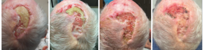
Rarely Basal Cell Carcinoma can be very aggressive infiltrating deeply into tissue. Some BCC’s can invade from skin into bone and cartilage. Some can even penetrate into vital structures such as ear canals and orbit. Although most BCC are visible to the skin, some can grow rapidly and extensively without showing much surface changes. Some rare BCC can be so deeply invasive and infiltrative that surgical treatment  is no longer possible. We have had experiences with BCC’s invading cranial cavity, orbit, and maxilla. Some of these require radical soft tissue and bone resection or orbital exenteration. Some BCC’s are simply not treatable surgically.

Related Articles

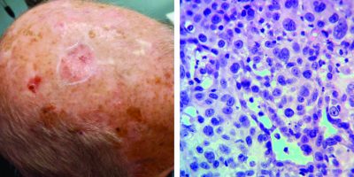
Management of Non-Healing Lesion
HISTORY An 85-year-old man with a history of multiple BCC and SCC of the scalp previously treated with radiation and intralesional 5-FU presents with a non healing lesion of the left parietal scalp. Shave biopsy of the left parietal scalp performed on 7/21/21 showed a non specific ulcer with atypical squamous epithelium of the scalp.…
Read More
A Case of Mistaken Identity: Porocarcinoma of the Scalp Initially Diagnosed as Basal Cell Carcinoma
History A 72-year-old man presented with a 2 year history of right anterior scalp lesion. The lesion was previously treated by an outside physician with cryosurgery multiple times over the course of 1 year. After a lack of resolution, the lesion was biopsied resulting in a diagnosis of basal cell carcinoma. No further treatment was…
Read More
Intralesional 5-fluorouracil for Cutaneous BCC and SCC
HISTORY  An 86-year-old man on hemodialysis with multiple medical problems and on warfarin presented with two carcinomas of the nasal tip: BCC of the left nasal tip and SCC of the right nasal tip. The ulceration of the right SCC exposed the alar cartilage. Patient completed six (6) 5-fluorouracil injections over a 5-week period. Complete…
Read More
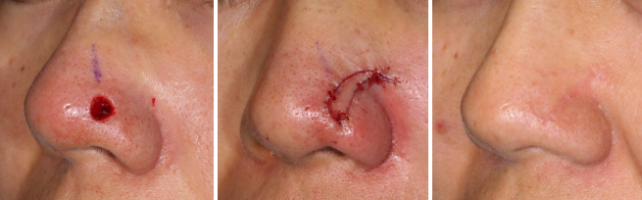
Nasal Open Wound Complication
HISTORY A 73-year-old man with BCC of the nasal tip presented 1 week following Mohs excision. His reconstruction was delayed for an additional week for a cardiac work up. Nasal reconstruction was performed with a superior extended nasal myocutaneous flap (SENMI flap) and nasal septal cartilage grafts. The patient developed nasal tip incisional discharge 1…
Read More
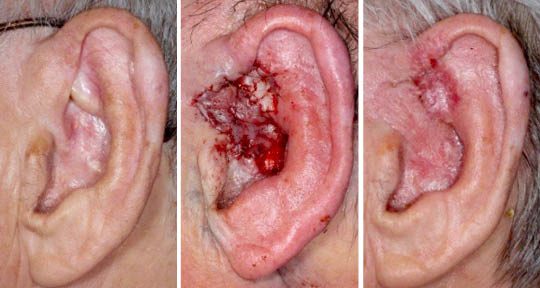
Recurrent BCC of the External Ear After Radiation
HISTORY A 77-year-old man presents with 5-year history of infiltrative basal cell carcinoma (BCC) of the left ear fossa triangularis and root of helix. The area was originally treated with four weeks of radiation in 2014, complicated by temporary ulceration and healing with adhesion. When the area developed crusting a couple of years later, the…
Read More
trichoepithelioma-histology-lateral-nasal-island-orange-county-skin-cancer-desmoplastic-series
Trichoepithelioma
HISTORY 33-year-old woman presents with 3-year history of slowly growing nasal tip lesion biopsied as a trichoepithelioma. Mohs excision was performed creating a 0.6 cm defect. It was reconstructed with a lateral nasal island flap. DISCUSSION Trichoepithelioma is a benign basaloid follicular neoplasm (arising from a pilosebaceous unit). Its basal cell proliferation is differentiated from…
Read More

STAY UP TO DATE

Receive research updates, inspiring stories, healthy living tips and more.

Something went wrong. Please check your entries and try again.

FOLLOW US

ELEVATING THE STANDARDS OF SKIN CANCER MANAGEMENT.

© 2018 SCARS Management Services. All rights reserved.