Dermatopathology
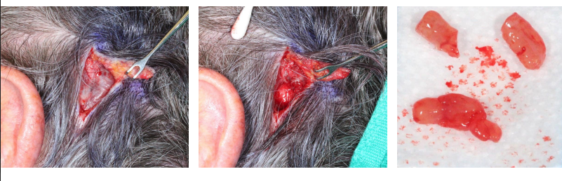
Subcutaneous Intravascular Pyogenic Granuloma of the Scalp of a POEMS Syndrome Patient
ABSTRACT An 82-year-old male with a past medical history of POEMS syndrome presented with a 6-weekhistory of a growing intravascular pyogenic granuloma in the subcutaneous scalp. Biopsyshowed lobular intravascular capillary hemangioma, also known as pyogenic granuloma. Lobularcapillary hemangiomas are most commonly found on the skin and represent some of thecutaneous changes seen within POEMS syndrome.…
Read More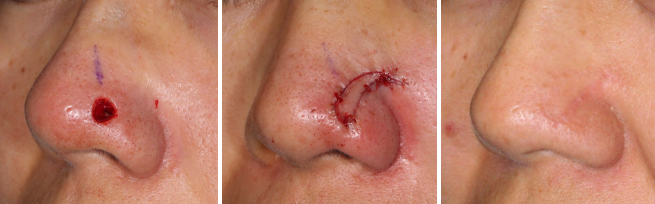
Trichoepithelioma
HISTORY 33-year-old woman presents with 3-year history of slowly growing nasal tip lesion biopsied as a trichoepithelioma. Mohs excision was performed creating a 0.6 cm defect. It was reconstructed with a lateral nasal island flap. DISCUSSION Trichoepithelioma is a benign basaloid follicular neoplasm (arising from a pilosebaceous unit). Its basal cell proliferation is differentiated from…
Read More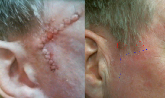
Recurrent Bowen’s Disease of Temple
HISTORY 63-year-old man presents with 9-month history of Bowen’s disease of right anterior and posterior sideburn biopsied on 08-20-18. History of previous Mohs excision of Bowen’s disease of the same area in February 2016. Patient has a history of multiple basal cell carcinomas, squamous cell carcinomas, and melanoma. DISCUSSION Management of this lesion is best…
Read More
Scleromyxedema Cutaneous Lesions
HISTORY 87-year-old man with underlying scleromyxedema and history of multiple basal cell carcinomas presents with a left earlobe nodule. Biopsy revealed this to be a lesion with benign spindle cell proliferation in vague concentric whorls with associated dermal fibrosis and chronic inflammation. This was a typical scleromyxedema lesion presenting within the fatty earlobe and not…
Read More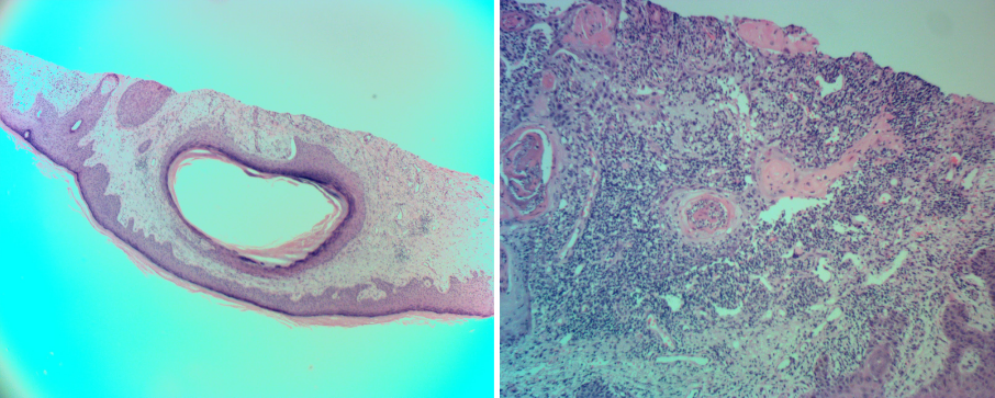
Cystic Squamous Cell Carcinoma of Forearm Skin
HISTORY 74-year-old man presents with 3-month history of painful lesion of the right forearm. Biopsy found cystic squamous cell carcinoma with deep margins involved. Wide local excision was performed achieving clear margins. DISCUSSION Cystic degeneration within squamous cell carcinoma of the skin is not considered an especially malignant finding. These histologic findings may represent changes…
Read More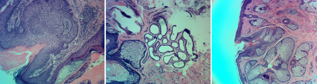
Sebaceous Nevus of Jadassohn
HISTORY 21-year-old male presents with 10-year history of left cheek lesion that has become more prominent with puberty and thickened over time. An excision found sebaceous nevus of Jadassohn. DISCUSSION – Nevus Sebaceous These congenital skin lesions are often excised. Timing and rationale of excision are based on the science behind the nevus sebaceous. Congenital…
Read More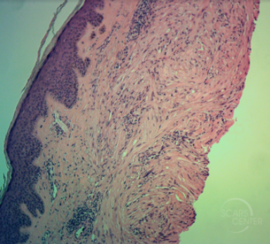
Leiomyoma of arm
HISTORY 74-year-old man presents with 0.6cm nodule on the posterior upper arm skin . Pathology showed atypical spindle cell neoplasm, favoring pleomorphic leiomyoma. Wide local excision was performed. DISCUSSION Leiomyoma is a tumor of smooth muscle cells. Leiomyoma of the skin usually arises from the arrector pili muscle of the hair follicles. The extent of…
Read More
Management of Recurrent Large Melanoma In Situ
HISTORY 78-year-old man presents with a recurrent melanoma in situ of left cheek in April 2018. Previously, the melanoma in situ was excised in 2001. Patient’s dermatologists performed excision in three stages over a period of 3 weeks to achieve clear margins. Reconstruction of left cheek was performed with a cheek and neck platysma myocutaneous…
Read More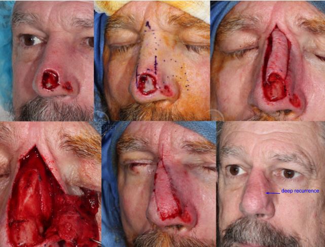
Recurrent SCC of Nasal Tip
HISTORY 71-year-old man presents with recurrence of SCC of nose following Mohs excision on 2-9-17. Partial rhinectomy was performed with incomplete Mohs excision leaving residual positive margins due to patient’s comfort. Two additional excisions of involved margins were done under anesthesia achieving clear margins. First stage reconstruction was performed on 4-11-18. DISCUSSION Management of a…
Read More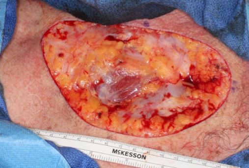
Upper Chest BCC PNI
HISTORY 53-year-old man presents with a 4-year-history of BCC of upper chest. The tumor measured 12 cm x 6 cm. Wide local excision with 1 cm margins on 11/10/2017 found clear margins and perineural invasion. The nerves up to 0.23 mm were involved by tumor. The final defect was 13.5 cm x 8.3 cm. …
Read More