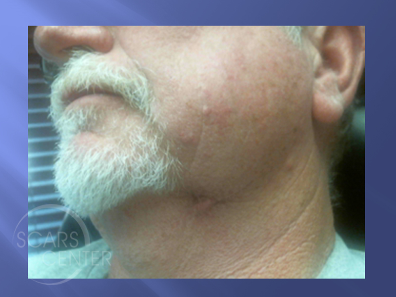
LEFT NECK METATYPICAL BASAL CELL CARCINOMA
HISTORY
63 year old man presented with a 5-year history of a lesion on the left anterior neck. The lesion was diagnosed as basal cell carcinoma and excised in 2012 , but it recurred three years ago with progressive deep scar growth. The lesion was re-biopsied in 2017 as a metatypical basal cell carcinoma with signet ring cell component associated with the old scar.
DISCUSSION
This is a case of a recurrent basal cell carcinoma in a 63-year-old. Originally, this man presented five years previously with left anterior neck lesion that was diagnosed as basal cell carcinoma and excised. Two years later, the patient noticed a small scar nodule deep within his old scar. The patient failed to realize the significance of that and only seeked evaluation as this presumptive scar grew over the next three years when the lesion was biopsied. Recently, it was found to be a metatypical basal cell carcinoma with signet ring cell component. This case was presented due to the unusual finding of signet ring cell component of this metatypical basal cell carcinoma.
This does not represent a more aggressive basal cell carcinoma based solely on the histology. Although some metatypical basal cell carcinomas can exhibit aggressive behavior, the metatypical features here likely associated with its location in the scar. The signet ring cell component itself has no particular significance for aggressive behavior. The patient with the deep scar recurrence in the neck requires evaluation for possible deeper invasion. Recurrent cancer deep within the scar can adhere to the deeper structures and deeper fascia including neurovascular tissue and underlying organs. In this patient, the concern was over the involvement of the submandibular gland, which was ruled out with the physical examination and an additional MRI. Bimanual finger palpation also was required to confirm easy mobility of this deep subcutaneous nodule from surrounding deep tissues.
The recommended treatment was Mohs excision. However, precaution must be taken in this particular area to avoid injury to the underlying facial artery. Due to the depth of the invasion of this cancer, involvement of the marginal mandibular branch of the facial nerve was likely. As a result, the patient needs to be counseled on the loss of the nerve function and asymmetry of the lower lip with smiling. Mohs surgery will be done in our center with the head and neck surgeon available to take an additional margin if proximity to the facial artery is a concern.

