Sebaceous Nevus of Jadassohn
HISTORY
21-year-old male presents with 10-year history of left cheek lesion that has become more prominent with puberty and thickened over time. An excision found sebaceous nevus of Jadassohn.
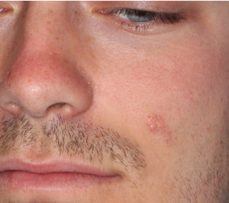
Fig. 1. Nevus sebaceous of the left cheek.
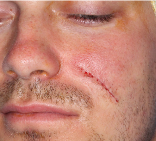
Fig. 2. Immediately after excision of the cheek lesion.
DISCUSSION – Nevus Sebaceous
These congenital skin lesions are often excised. Timing and rationale of excision are based on the science behind the nevus sebaceous.
- Congenital epidermal hamartoma
- Responds to hormonal influences
- From hypoplastic before puberty to hyperkeratosis and papillomatosis with numerous and hyperplastic sebaceous glands after puberty
- clinically the lesion thickens with age
- Most commonly presents in the head and neck – 95%
- Neoplastic transformation occurs uncommonly
- Most common tumors arising within are benign
- syringocystadenoma papilliferum
- trichoblastoma (may be misdiagnosed as Basal Cell Carcinoma)
- BCC is the most common malignant transformation (deletions of patched gene)
- SCC, adnexal carcinomas, malignant melanomas have been reported
- 5-20% lifetime risk of malignant degeneration (higher incidence studies had trichoblastomas misdiagnosed as BCC?)
- 0.8% of excised sebaceous nevi in adolescents had BCC in one study (misdiagnosed trichoblastomas?)
- Long duration associated with malignant change
- Most common tumors arising within are benign
- Consider excision after puberty
- When large or with Epidermal Nevus Syndrome (Jadassohn Nevus Phakomatosis)
- CNS problems – Developmental delay, seizures, intracranial mass
- Bone problems – bone hypertrophy, spina bifida
- Eye problems – optic nerve hypoplasia, oculomotor dysfunction
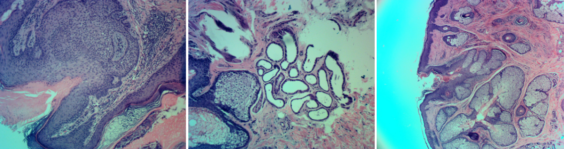
Fig. 3. Histomicrographs of nevus sebaceous.