Mapping of Recurrent Melanoma In Situ of Leg
HISTORY
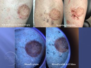
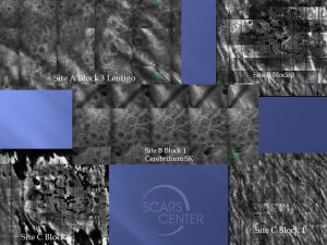
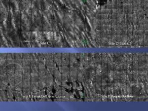
50-year-old woman presents with recurrent melanoma in situ of left leg. First biopsied on 9-2-16, the lesion was excised on 10-14-16 and showed extensive residual melanoma in-situ with radial margins involved from 12- 3 o’clock. The defect was reconstructed at the time with a full thickness skin graft. Second excision cleared the involved margins on 11-7-16. A new pigmented lesion occurred at the 7 o’clock excision margin 1 year later. The biopsy on 9-20-17 showed severely atypical lentiginous junctional melanocytic proliferation. Left leg additional margin excision on 10-24-17 showed extensive recurrent melanoma in situ. Patient has family history of melanoma in her father. The patient underwent extensive mapping with Wood’s lamp, laser reflectance confocal microscopy, and mapping punch biopsies.
DISCUSSION
This recurrent melanoma in situ of the leg has poorly defined margins. Instead of chasing the margins with serial excisions, we planned the excision with noninvasive imaging guided punch biopsies. The noninvasive imaging included a Wood’s lamp and reflectance confocal microscopy. After clear excision margins are confirmed, the area will be treated with 12 weeks of imiquimod. This topical mop-up of residual cells is in the effort of avoiding the 17-20% recurrence rate after excision of melanoma in situ.


