RECURRENT SCALP MELANOMA WITH CALVARIAL METASTASES
HISTORY
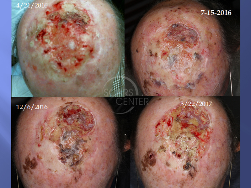
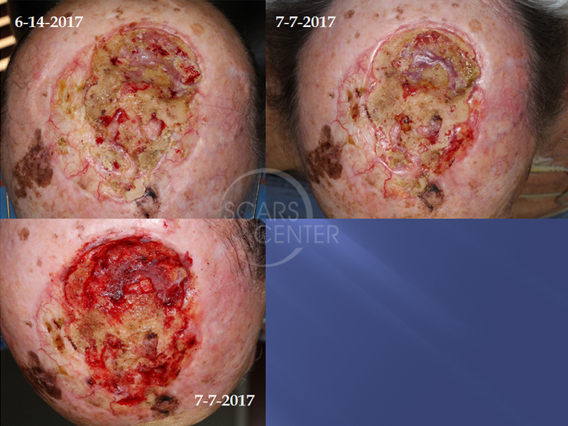
85-year-old man presented in 4/2016 with a 4 month history of a nonhealing scalp wound following excision of melanoma done in 2014, then additional excision with skin graft closure followed by radiation completed in June 2015. The wound developed skin breakdown and osteoradionecrosis treated with serial outer table of calvarium debridements.
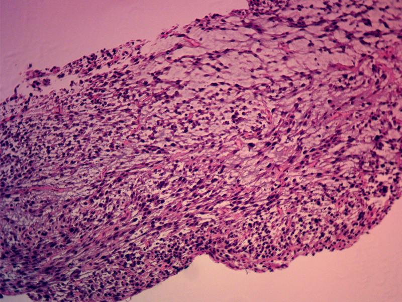
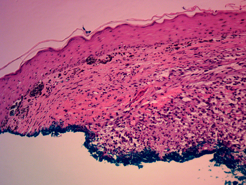
Recent biopsy on 8/1/2017 of left superior central forehead showed malignant melanoma with myxoid spindle cell features.
DISCUSSION
The myxoid spindle cell features of this suspected melanoma is an unusual finding. Additional histologic consultation with additional staining is required to differentiate this tumor from a possible sarcoma variant. Ronald Bar, M.D. recommended consultation with Sharon Weiss, M.D. at Emery University who is a national expert on soft tissue tumors.
We reviewed the original melanoma from 2013. The histology and architecture of this tumor was significantly different from the new suspected melanoma. A suggestion was made that this new myxoid spindle cell / melanoma lesion may be a radiation induced sarcoma. That is possible, but unlikely given the two-year span since completion of radiation. Radiation induced sarcomas usually follow at least five years afterwards or longer.
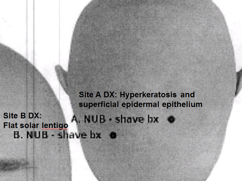
This patient will require evaluation by an oncologist with a likely PET scan for staging. It is interesting to follow the serial photographs of this patient’s left upper forehead lesion. As can be seen in the photos over a period of one and a half years, there is progressive increase in pigmentation. Biopsy of this pigmentary change a year ago was benign. The latest change diagnosed as melanoma was a nodule.

