Dysplasia of Lower Lip
HISTORY
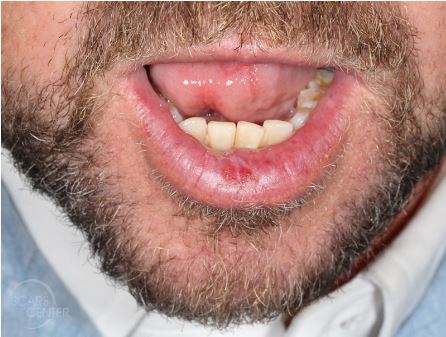
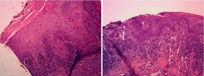
51-year-old man presents with one-year history of lower lip nonhealing lesion. The patient has a many year history of extensive sun exposure with scaling and crusting. A biopsy of the nonhealing lesion showed leukokeratosis with minimal keratinocytic atypia. Excisional biopsy done 11-1-17 showed moderate to focally severe squamous atypia. The patient was then treated with photodynamic therapy of the lower lip. Levulan Kerastick (aminolevulinic acid) was applied, followed by 4 hours of incubation and blue light therapy.

Chronically inflamed lower lip lesion.

Moderate to focally severe squamous atypia of lower lip.
DISCUSSION
The presentation of a nonhealing lesion within actinic cheilitis of the lower lip demands definitive diagnosis to rule out invasive squamous cell carcinoma. Shave or punch biopsy of the lesion is a good start towards identifying the diagnosis. However, this approach can miss invasive carcinoma. Actinic cheilitis lesions tend to exhibit heterogeneous findings within a single lesion – from keratosis to atypia to carcinoma in situ to invasive squamous cell carcinoma. Complete excision of the lesion provides that definitive diagnosis and complete lesion evaluation. In addition, it serves to resolve the recurrent symptoms of bleeding and crusting of the lesion.
To treat the patient’s underlying actinic cheilitis, the patient was involved in the decision. Several available options can be divided into the “slow and crusty” and “fast and furious” approaches. The “slow and crusty” include topical chemotherapy with 5-fluorouracil. The “fast and furious” include laser resurfacing, TCA chemical peel, or photodynamic therapy (PDT). We chose photodynamic therapy.
Although, lip shave with mucosal advancement has been described for actinic cheilitis, it is rarely employed by us at the SCARS Center. It is a very aggressive technique removing most of the lower red lip mucosa that is reserved for special high risk cases. We prefer excisions directed at specific lesions as described here.
The patient was treated with a 4 hour incubation time which is an intermediate incubation time for PDT overall but ranges on the aggressive for the thin skin of the lip. The response was robust with 2 days of redness and swelling followed by 5 days of bleeding and crusting, then another 3 days of light ulceration and scaling.
