Archive for March 2011
Basosquamous Cell Carcinoma
Metatypical basosquamous cell carcinoma is a subset of cutaneous cancers that poses an increased risk of metastases to the patient due to its aggressive behavior. The classic histology of metatypical basosquamous cell carcinoma includes features of both basal cell carcinoma and squamous cell carcinoma, as well as areas of intermediate differentiation. The cells are larger and…
Read MoreNew Melanoma Staining: SOX10 and MiTF
S-100, tyrosinase, Melan A, and HMB 45 have been the workhorses of melanocyte histopathology. A new immunohistochemical stain, SOX10, has recently been shown to be a useful marker in the diagnosis of melanocytic tumors. It is strongly expressed by desmoplastic melanoma and is less likely to be expressed by background fibrocytes and histiocytes. Sry-related HMG-BOX…
Read MoreRadiation Therapy of Skin Cancer: Dosing and Complications
Although, radiation of ear carcinomas can pose a risk of chondritis and poor healing, that risk can be virtually eliminated with lower dose fractions (radiation dose per treatment) spread over a longer course. Total dose requirement for skin cancer treatment ranges from 3500 cGy to 6000 cGy depending on the size and depth of the…
Read MoreSquamous Cell Carcinoma Lower Lip
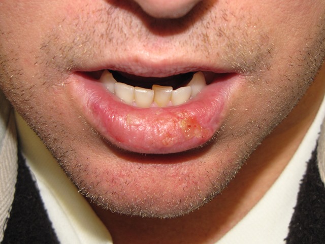
A man in his thirties with lifelong h/o severe actinic cheilitis of left lower lip. Recurrent flare-ups of cheilitis have been treated as recurrent herpetic infection. Recently he has experienced worsening of cheilitis and appearance of the lesion. Biopsy found squamous cell carcinoma with perineural invasion. Lip invasion was histologically measured to be > 2mm…
Read MoreAcceptance of Residual Melanoma-In-Situ: Avoiding Cosmetic and Functional Deformation
Acceptance of residual melanoma-in-situ (lentigo maligna) after an excision can avoid cosmetic and functional deformation. Is the residual risk of recurrence and transformation into invasive melanoma low enough to warrant observation? Melanoma-in-situ (MIS) is a high risk lesion due to three primary reasons. The first is that a partially biopsied melanocytic lesion with a diagnosis…
Read MoreMelanoma-in-situ of the Chin
A middle-age man with previous melanoma-in-situ of the back presents with new chin lesion biopsied as melanoma-in-situ on a deep shave biopsy. Subsequent surgical excision with 2 mm margins revealed no evidence of residual melanoma. Observation was chosen as the course of management. Should additional wider margin resection be performed? Melanoma-in-situ (MIS) is a high…
Read MorePerineural Invasion
Perineural invasion is strongly indicative of aggressive behavior and risk of metastases. True perineural invasion requires histologic finding of cancer invasion of the nerve or cancer invasion of the epineurium adjacent to the nerve. This must be distinguished from cancer near a nerve, cancer encompassing a nerve, and perineural inflammation. Cancer cells within the nerve…
Read More

