Archive for September 2017
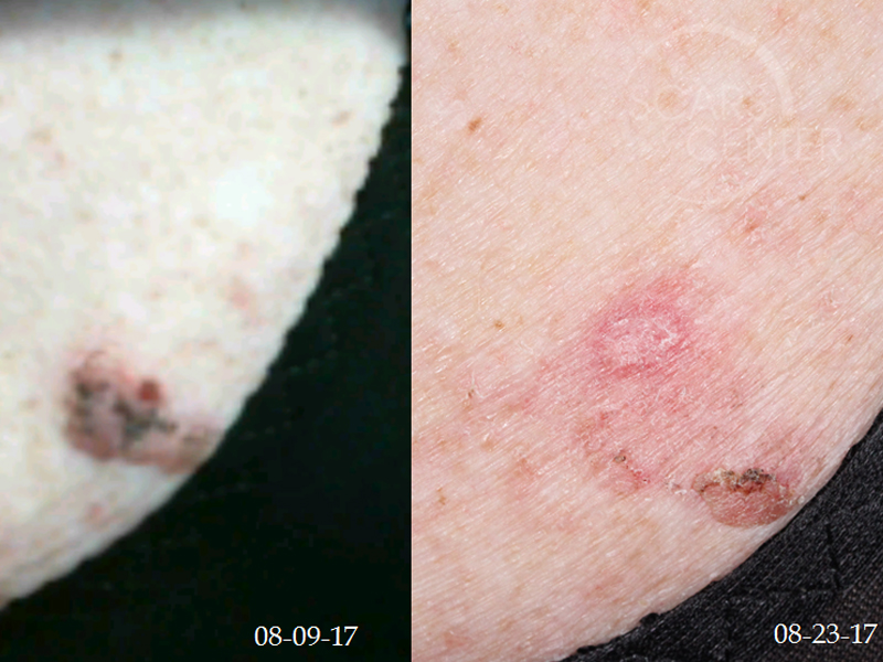
INFLAMED SK APPEARING AS SQUAMOUS CELL CARCINOMA
HISTORY 57-year-old woman with 40-year history of growth on right mid back that has changed in color and size. This is a 2.2cm rough plaque with variable colors. Previous biopsy done 15-20 years ago was benign. Patient has a history of BCC and SCC. Biopsy was read as a squamous cell carcinoma by a dermatologist.…
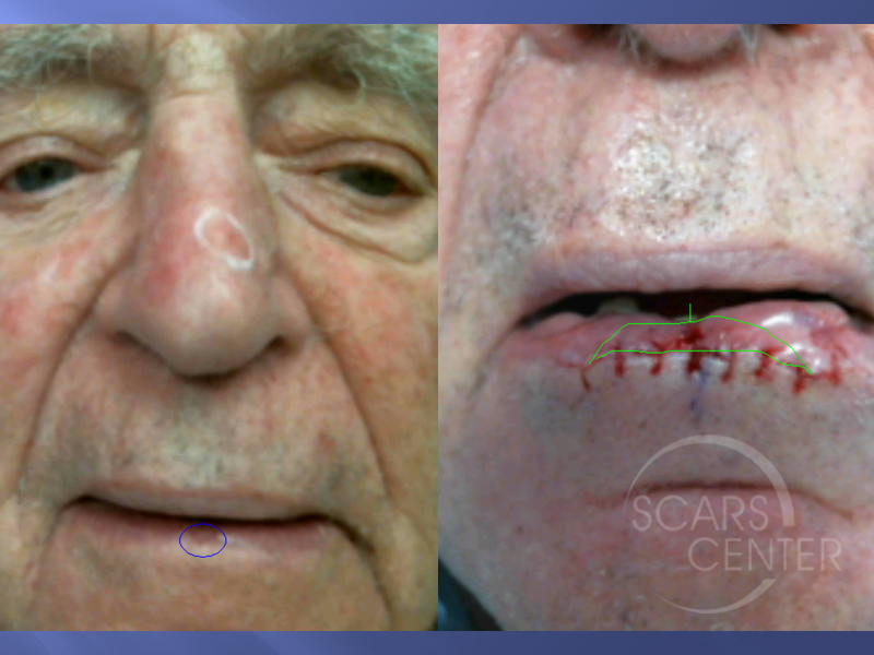
Read MoreLOWER LIP ATYPIA AT MARGINS OF SQUAMOUS CELL CARCINOMA
HISTORY 89-year-old man presents with several month history of lesion on bottom lip. A biopsy was taken on 8-9-17 and showed severe squamous atypia without ruling out deeper SCC. Excision of lesion done 9-6-17 showed invasive squamous cell carcinoma with foci of moderate squamous dysplasia from 9 to 3 o’clock. Patient has history of…
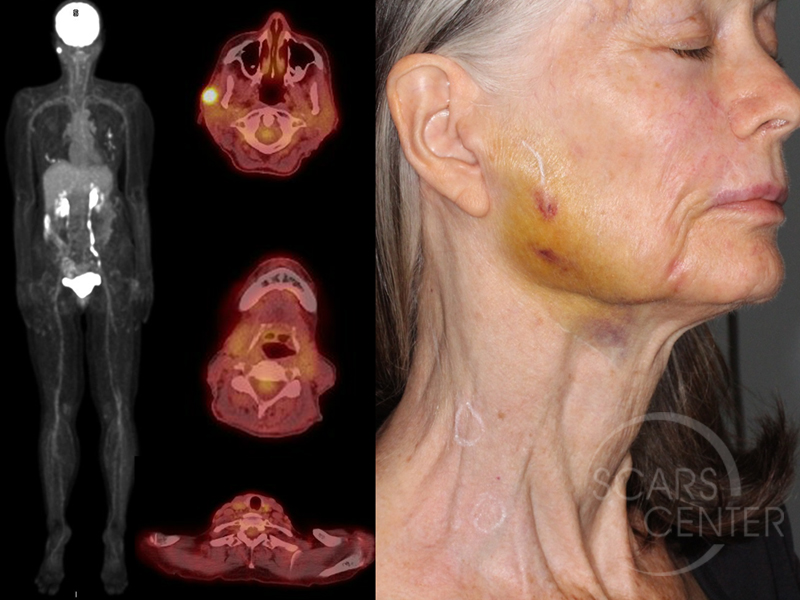
Read MoreRECURRENT MELANOMA
HISTORY 70-year-old woman presents with metastatic melanoma in the right parotid gland region of 1 year duration. Diagnosis was made with a core needle biopsy on 09-08-17. Patient also has small palpable right cervical lymph nodes with SUV 1.6 and 2.0. Patient history of melanoma began in 2001 as a pink macule of the…
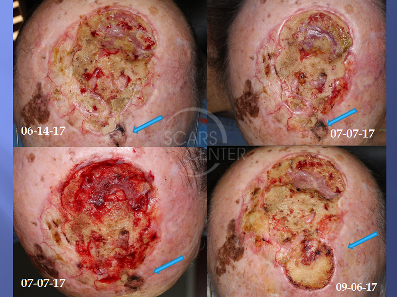
Read MoreRECURRENT SCALP MELANOMA WITH CALVARIAL METASTASES
HISTORY 85-year-old man with history of treated scalp melanoma presented with a 1 month rapidly growing scalp nodule. The original melanoma was treated in 2014 with excision, then re-excision of recurrence, and post-operative radiation in June 2015. The wound developed skin breakdown and osteoradionecrosis and was treated with serial outer table of calvarium debridement. Biopsy…
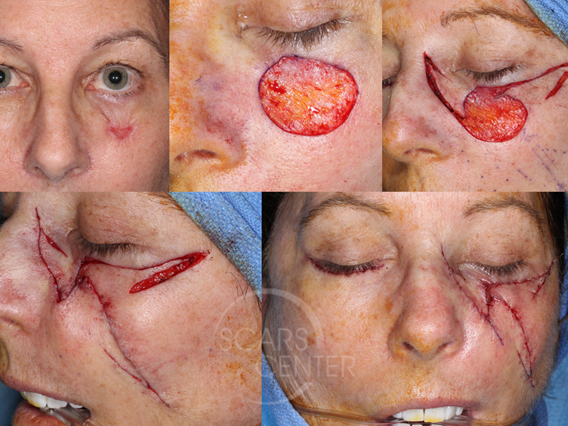
Read MoreCHEEK AND EYELID RECONSTRUCTION WITH MULTIPLE PARTIAL ISLAND FLAPS
HISTORY 61-year-old woman presents with several year history of pigmented left cheek and lower lid lesion. She was initially treated with liquid nitrogen 15 months ago. A biopsy was performed 6-8-17 and revealed melanoma in situ. Excision with close margins performed 6-16-17. Clear margins found on LPMG reading. Second opinion from UCSD was suspicious for…
Read MoreMULTIPLE ATYPICAL NEVI WITH STRONG FAMILY HISTORY OF CANCER
HISTORY 24-year-old woman presents with multiple atypical nevi. Almost all lesions had mild atypia, few had focally moderate atypia, and one on her right posterior shoulder had severe atypia and was excised with 5mm margins elsewhere. Family history is significant for melanoma in grandmother and pancreatic cancer in her father. Physical exam showed multiple 2-4…
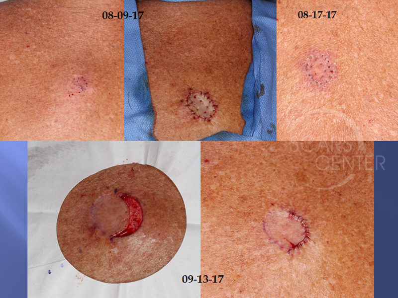
Read MoreADDITIONAL MARGIN EXCISION AFTER SKIN GRAFTING
DISCUSSION This series of photographs represents an approach to margin management of melanoma in situ. Some melanoma in situ margins are difficult to assess clinically, as is the case in our first series with extensively pigmented skin. In some settings, taking 7-9 mm margins exposes the patient to significant healing issues, as the second case…
Read More
