RECURRENT SCALP MELANOMA WITH CALVARIAL METASTASES
HISTORY
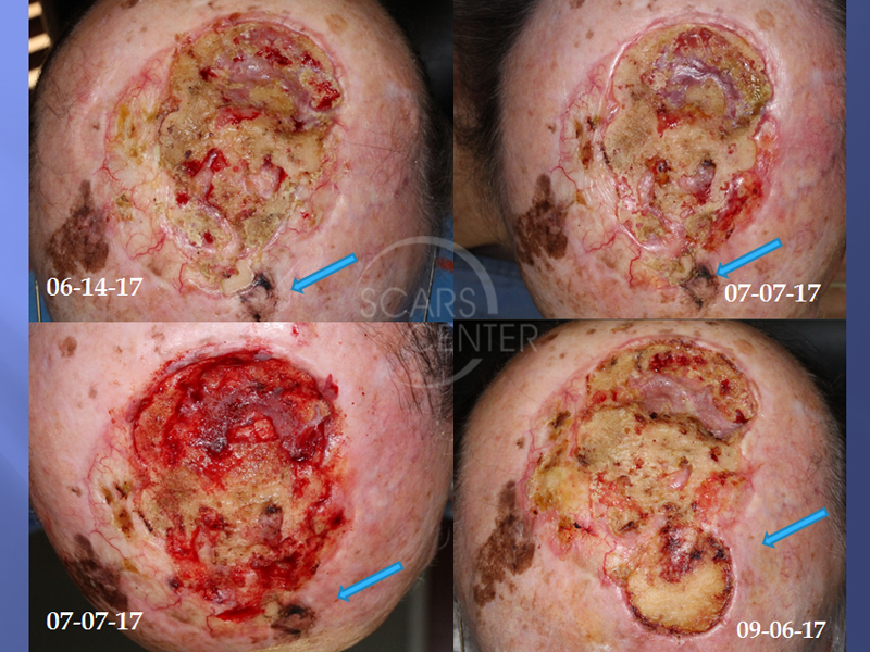
85-year-old man with history of treated scalp melanoma presented with a 1 month rapidly growing scalp nodule. The original melanoma was treated in 2014 with excision, then re-excision of recurrence, and post-operative radiation in June 2015. The wound developed skin breakdown and osteoradionecrosis and was treated with serial outer table of calvarium debridement.
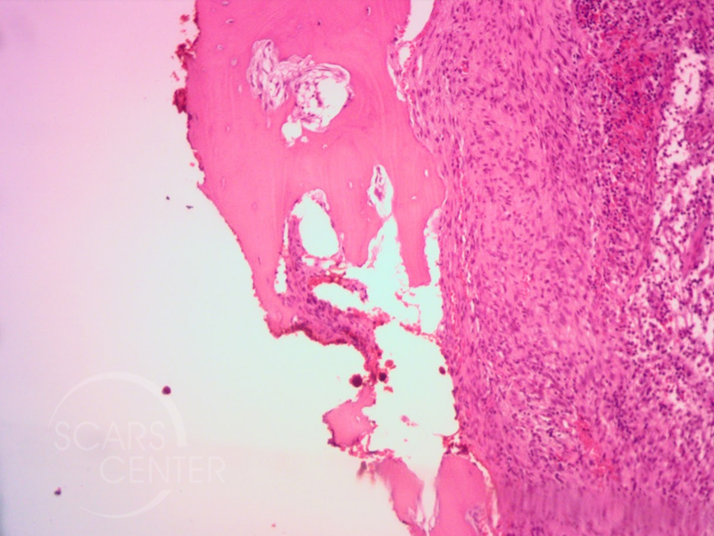
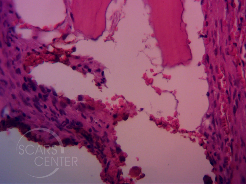
Biopsy on 8-1-2017 of left forehead rapidly growing nodule showed malignant melanoma with myxoid spindle cell features. The lesion was located adjacent to the surgical field of original melanoma and within the radiation field. Wide local excision with underling bone cortex on 8-28-2017 showed extensive residual malignant melanoma and myxoid spindle cell proliferation invading into bone. This was present in the setting of malignant melanoma, lentigo maligna type. The tumor was judged to be a dedifferentiated melanoma. (see histomicrographs)
DISCUSSION
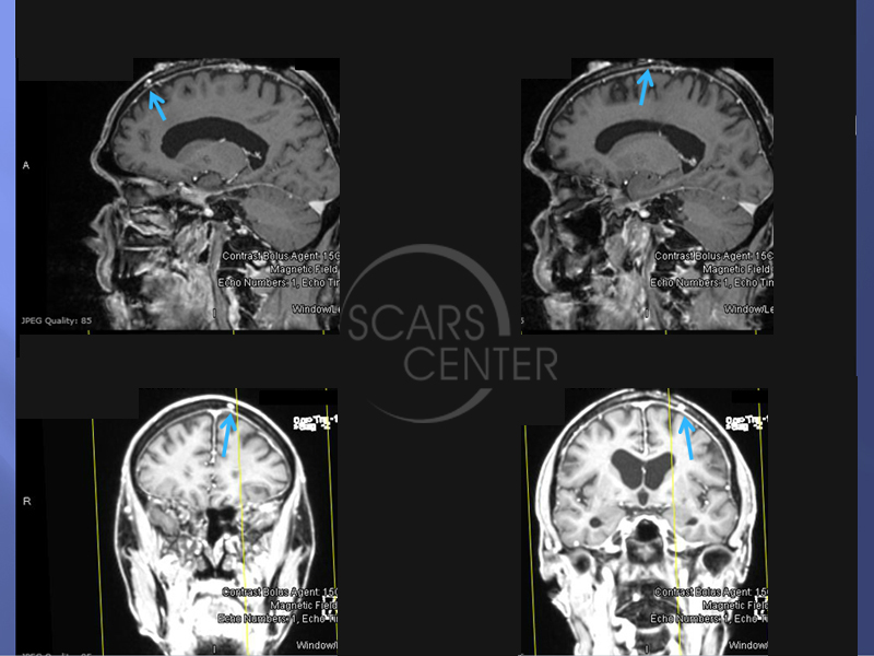
MRI of the head found two calvarial metastases within 2 cm of the tumor in the diploic space of the calvarium. (See MR images)
The diagnosis of melanoma offers the patient an opportunity to be treated with Keytruda (pembrolizumab). This biological / antibody targets PD-1 pathway. It is indicated in several solid tumors including metastatic melanoma, non-small cell lung cancer, head and neck cancers, Hodgkin’s lymphoma, and urothelial cancers.

