Atypical Nevus – a Melanoma Precursor? Not Exactly
HISTORY
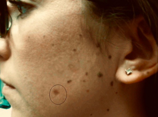
A 27-year-old woman presented with many-year history of left cheek nevus. The lesion grew in size and changed in color. Shave biopsy showed a nevus with moderate atypical melanocytic hyperplasia. Residual pigmentation of the nevus and involved histologic margins remained after the biopsy.
DISCUSSION
Are all atypical melanocytic lesions pre-melanomas? Should dysplastic lesions be promptly excised to avoid development of melanoma? Signs of melanoma and melanoma risks have been broadly publicized in the media and are widely known by the public. The increased anxiety over melanoma risks has translated into more frequent biopsies and more aggressive surgical management. But a new multi-institutional study dispels that notion, at least for atypical nevi.
These dysplastic moles have been treated aggressively with a 2 mm margin excision. New data supports observation of the previously biopsied moles instead of excision. The study published in JAMA Dermatology in October 2018 addressed moderately atypical lesions.* The retrospective study of previously biopsied dysplastic nevi found that none of them transformed into a melanoma over an average 7 year follow-up.
The fact is that the patients with dysplastic nevi are at an increased lifetime risk of melanoma. However, melanoma anxiety should not focus on the lesions confirmed as atypical. Instead of chasing each atypical mole with complete excision, the patient’s entire skin needs to be regularly examined. Conclusion? Less scars by surgeons and more self exams by patients.
There is a catch to these recommendations. One needs to be certain that the mole is indeed only moderately atypical. Partial biopsies leaving pigmented portions of mole do not definitively rule out more aggressive components of a mole. Given the heterogeneity within an atypical melanocytic lesion, these more aggressive components need to be ruled out with complete sampling. Therefore, a virtually complete shave biopsy with positive histologic margins does not need additional treatment. Residual or recurrent pigmentation requires additional biopsy or excision to rule out more aggressive components.
*Kim CC, Berry EG, Marchetti MA, et al. Risk of subsequent cutaneous melanoma in moderately dysplastic nevi excisionally biopsied but with positive histologic margins. JAMA Dermatol. 2018;154(12):1401-1408.