Posts Tagged ‘skin graft’
Nasal Skin Graft Revision with Flaps
HISTORY The patient presented for scar revision 8 months after Mohs excision of BCC of the nose and closure with an ear skin graft, performed elsewhere. Patient presents for correction of the atrophic erythematous skin graft scar. The patient was treated with two signature flaps of the SCARS Center developed for nasal reconstruction. DISCUSSION The…
Read MoreSubtotal Ear Reconstruction with Alloplast
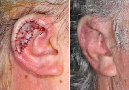
DISCUSSION Subtotal ear reconstruction is a challenging surgical task that often requires non-traditional techniques. This particular patient was treated with an alloplastic implant and rib cartilage grafts. We utilized Medpor Helical Rim implant, a porous polyethylene material (Stryker, USA) used for microtia reconstruction. The implant was used for its elegantly curved helix. The rib cartilage…
Read MoreMelanoma within recurrent MIS
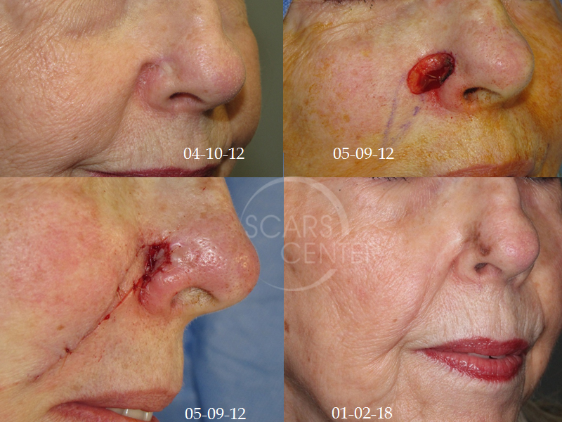
HISTORY 73-year-old woman presents with 1-year history of recurrent pigmented lesion of the right nasal ala found to be invasive melanoma with 0.5 mm invasion. Patient’s history began 9 years ago in 2009 with excision of a melanoma in situ and skin graft closure. In 2012, pigmentation adjacent to the graft site was found…
Read MoreLeft Ear Multiple Recurrent Basal Cell Carcinoma
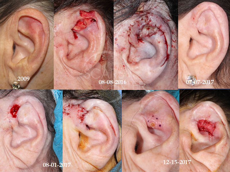
HISTORY 64-year-old woman presents with recurrence of left ear basal cell carcinoma. She has a history of sun lamp tanning and scuba diving with multiple sun burns. Basal cell carcinoma was first removed from left scapha and treated with a skin graft in July 2009. The area subsequently developed crusting and was treated with…
Read MoreHand SCC In Situ
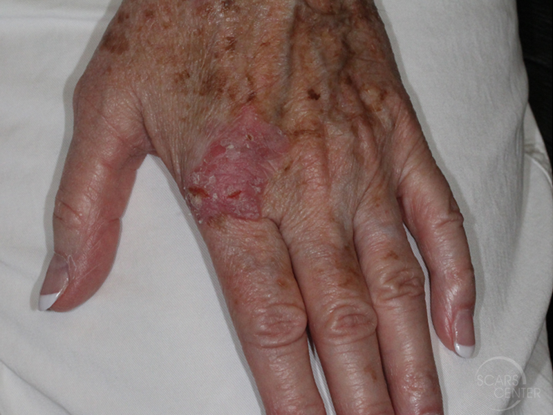
HISTORY 68-year-old woman presents with a many month history of squamous cell in situ of left hand. This large 2.5 cm lesion on the hand poses a unique challenge to ensuring hand function post-operatively. Treatment options include C&D (curettage and desiccation), curettage only with post-treatment imiquimod, Mohs excision with a skin graft closure, or superficial…
Read MoreADDITIONAL MARGIN EXCISION AFTER SKIN GRAFTING
DISCUSSION This series of photographs represents an approach to margin management of melanoma in situ. Some melanoma in situ margins are difficult to assess clinically, as is the case in our first series with extensively pigmented skin. In some settings, taking 7-9 mm margins exposes the patient to significant healing issues, as the second case…
Read MoreMELANOMA IN SITU OF BACK
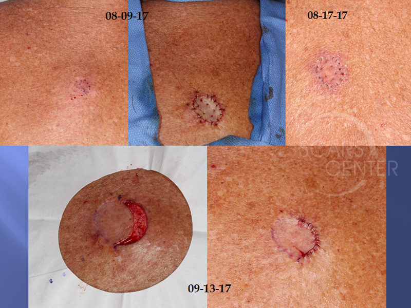
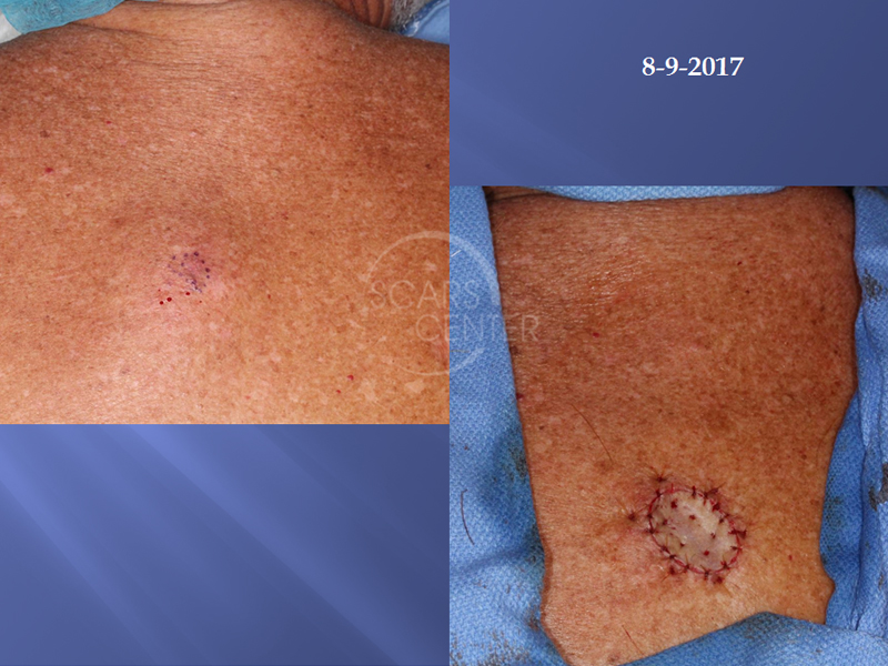
HISTORY 69-year-old man presents with melanoma in situ of the upper back. A biopsy on 6/15/2017 showed malignant melanoma at least in-situ and at least Clark’s level I with margins involved. Excision and skin graft closure performed on 8/9/2017. Pathology showed residual malignant melanoma in situ with involved margin at 2-4 o’clock. DISCUSSION Management…
Read More

