Melanoma
DNA Molecular Testing and Personalized Medicine – Part 2
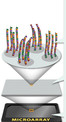
Another genetic molecular technology that has evolved tremendously is real time polymerase chain reaction (PCR). When accompanied by reverse transcription prior to PCR (RT-PCR), it has also allowed rapid multiple gene analysis useful for cancer staging such as OncotypeDx. This platform analyzes RNA for 21 genes with RT-PCR to stratify breast cancer patients according to…
Read More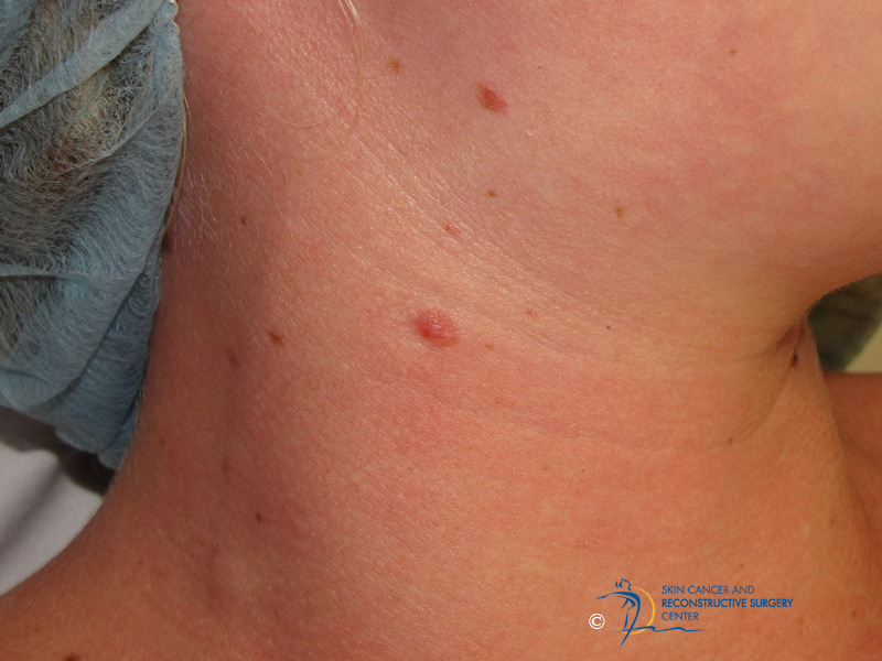
STUMP and MelTUMP
Ambiguous melanocytic lesions – those with histopathologic findings without a diagnostic consensus – are divided into thin and thick tumors. Thin lesions appear as dysplastic nevi and have a potential of being part of melanoma in situ or superficial spreading melanoma. We have explored diagnostic and therapeutic considerations of the thin lesions in a previous…
Read More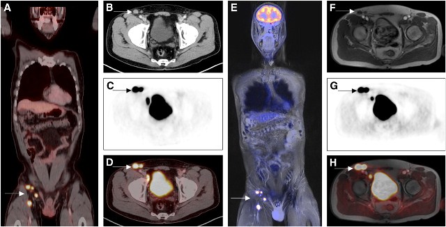
PET Imaging – Realizing a Star Trek Tricorder
PET scanning is utilized in skin cancer staging by detecting regional lymph node metastases as well as distant organ metastases. Its specificity is superior to CT and MR for N-staging, particularly with melanoma. It can be the superior modality for primary tumor staging as well. PET scan imaging is based on the increased metabolic activity…
Read More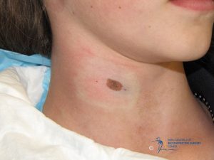
Atypical Nevus – Chasing Excision Margins
A 12 year old with a 10 x 7 mm pigmented nevus present since infancy undergoes excisional biopsy of the nevus for cosmetic and diagnostic purposes. Pathology evaluation finds this to be a compound nevus with moderate atypia and partial margin involvement. Pathologist recommends re-excision with 2 mm margins. Do we chase the margins as…
Read MoreNew Melanoma Staining: SOX10 and MiTF
S-100, tyrosinase, Melan A, and HMB 45 have been the workhorses of melanocyte histopathology. A new immunohistochemical stain, SOX10, has recently been shown to be a useful marker in the diagnosis of melanocytic tumors. It is strongly expressed by desmoplastic melanoma and is less likely to be expressed by background fibrocytes and histiocytes. Sry-related HMG-BOX…
Read More
Acceptance of Residual Melanoma-In-Situ: Avoiding Cosmetic and Functional Deformation
Acceptance of residual melanoma-in-situ (lentigo maligna) after an excision can avoid cosmetic and functional deformation. Is the residual risk of recurrence and transformation into invasive melanoma low enough to warrant observation? Melanoma-in-situ (MIS) is a high risk lesion due to three primary reasons. The first is that a partially biopsied melanocytic lesion with a diagnosis…
Read MoreMelanoma-in-situ of the Chin
A middle-age man with previous melanoma-in-situ of the back presents with new chin lesion biopsied as melanoma-in-situ on a deep shave biopsy. Subsequent surgical excision with 2 mm margins revealed no evidence of residual melanoma. Observation was chosen as the course of management. Should additional wider margin resection be performed? Melanoma-in-situ (MIS) is a high…
Read More