Patient Case Studies
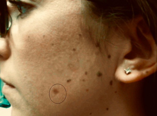
Atypical Nevus – a Melanoma Precursor? Not Exactly
HISTORY A 27-year-old woman presented with many-year history of left cheek nevus. The lesion grew in size and changed in color. Shave biopsy showed a nevus with moderate atypical melanocytic hyperplasia. Residual pigmentation of the nevus and involved histologic margins remained after the biopsy. DISCUSSION Are all atypical melanocytic lesions pre-melanomas? Should dysplastic lesions be…
Read More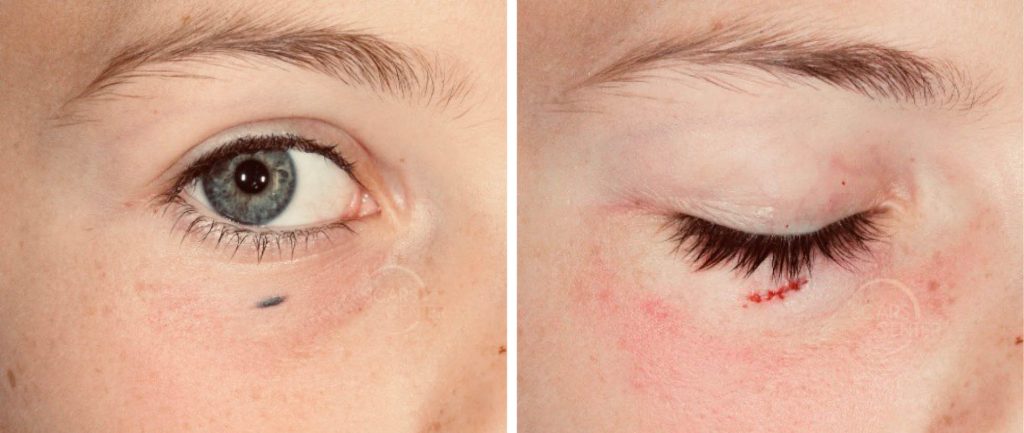
Pediatric Office Procedures with PRO-NOX™ Nitrous Oxide
HISTORY An 8-year-old girl presents with a 1 year history of dark mole of the right lower eyelid. Initially presenting as a small freckle, it grew and darkened in color. Right lower eyelid excisional biopsy was performed utilizing PRO-NOX™ nitrous oxide system. Biopsy showed intradermal nevus, combined type (primarily a common blue nevus and a…
Read More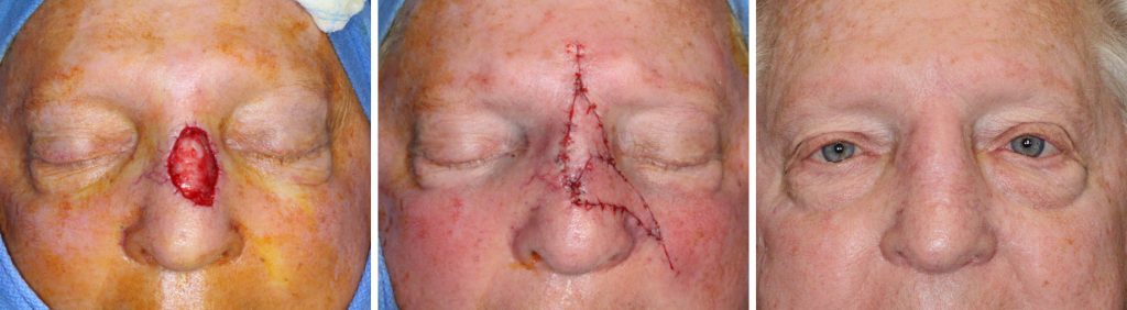
Nasal Skin Graft Revision with Flaps
HISTORY The patient presented for scar revision 8 months after Mohs excision of BCC of the nose and closure with an ear skin graft, performed elsewhere. Patient presents for correction of the atrophic erythematous skin graft scar. The patient was treated with two signature flaps of the SCARS Center developed for nasal reconstruction. DISCUSSION The…
Read More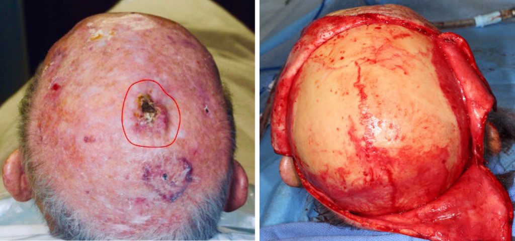
Leiomyosarcoma of Scalp
HISTORY An 84-year-old man presented with a 7-month history of 2.5 cm ulcerated nodule of the scalp. Biopsy showed a poorly defined neoplasm consistent with a leiomyosarcoma. Wide local resection with 1 cm margins cleared the tumor. The 5 cm scalp defect was reconstructed with multiple large scalp rotation flaps. DISCUSSION Spindle cell neoplasms of…
Read More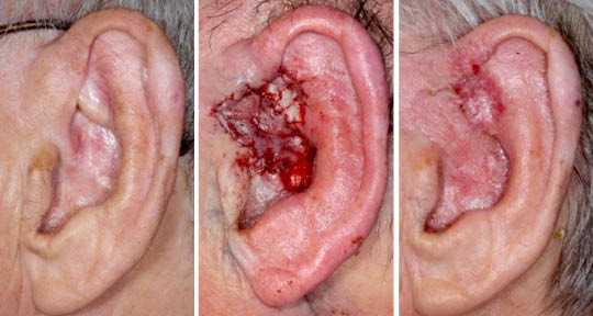
Recurrent BCC of the External Ear After Radiation
HISTORY A 77-year-old man presents with 5-year history of infiltrative basal cell carcinoma (BCC) of the left ear fossa triangularis and root of helix. The area was originally treated with four weeks of radiation in 2014, complicated by temporary ulceration and healing with adhesion. When the area developed crusting a couple of years later, the…
Read More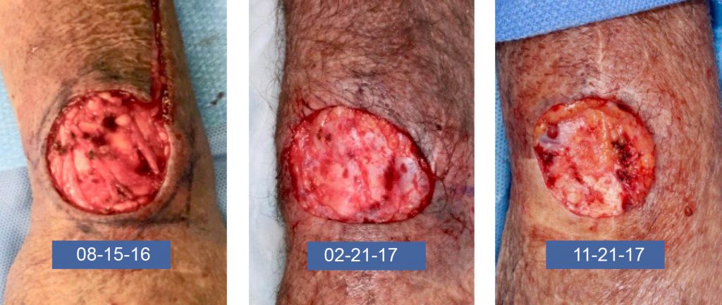
Lethal Squamous Cell Carcinoma of the Forearm in a Lung Transplant Recipient
HISTORY A 71-year-old lung transplant recipient presented with a recurrence of left dorsal forearm and wrist Squamous Cell Carcinoma (SCC) after Mohs excision in 2016 performed elsewhere. The patient underwent Mohs excision with clear margins and additional deeper wide local resection with no carcinoma identified. Patient was referred for radiation oncology evaluation, but he was…
Read More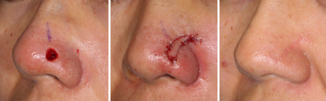
Trichoepithelioma
HISTORY 33-year-old woman presents with 3-year history of slowly growing nasal tip lesion biopsied as a trichoepithelioma. Mohs excision was performed creating a 0.6 cm defect. It was reconstructed with a lateral nasal island flap. DISCUSSION Trichoepithelioma is a benign basaloid follicular neoplasm (arising from a pilosebaceous unit). Its basal cell proliferation is differentiated from…
Read More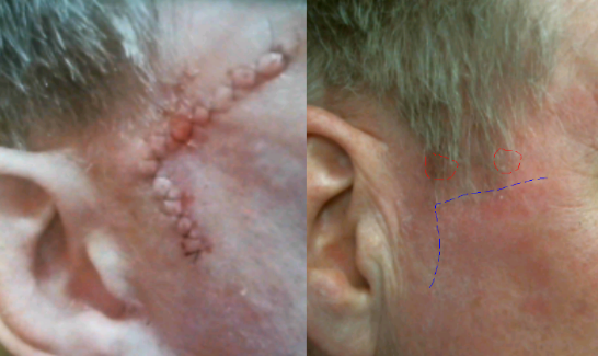
Recurrent Bowen’s Disease of Temple
HISTORY 63-year-old man presents with 9-month history of Bowen’s disease of right anterior and posterior sideburn biopsied on 08-20-18. History of previous Mohs excision of Bowen’s disease of the same area in February 2016. Patient has a history of multiple basal cell carcinomas, squamous cell carcinomas, and melanoma. DISCUSSION Management of this lesion is best…
Read More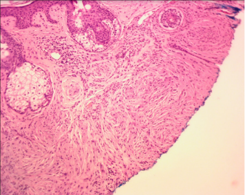
Scleromyxedema Cutaneous Lesions
HISTORY 87-year-old man with underlying scleromyxedema and history of multiple basal cell carcinomas presents with a left earlobe nodule. Biopsy revealed this to be a lesion with benign spindle cell proliferation in vague concentric whorls with associated dermal fibrosis and chronic inflammation. This was a typical scleromyxedema lesion presenting within the fatty earlobe and not…
Read More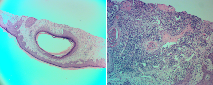
Cystic Squamous Cell Carcinoma of Forearm Skin
HISTORY 74-year-old man presents with 3-month history of painful lesion of the right forearm. Biopsy found cystic squamous cell carcinoma with deep margins involved. Wide local excision was performed achieving clear margins. DISCUSSION Cystic degeneration within squamous cell carcinoma of the skin is not considered an especially malignant finding. These histologic findings may represent changes…
Read More