Myxoid Lipoma of Scalp
HISTORY
61-year-old man presents with 40 year history of 2.5 cm mobile lipoma on left scalp vertex. Ultrasound performed on 11-14-17 revealed lipoma. Results from excision of mass showed myxoid lipoma. Consultation was obtained with Emory’s Sharon Weiss, MD, a soft tissue pathology expert.
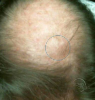
Soft tissue mass of the scalp. The mass inside the capsule appeared clear and gelatinous.
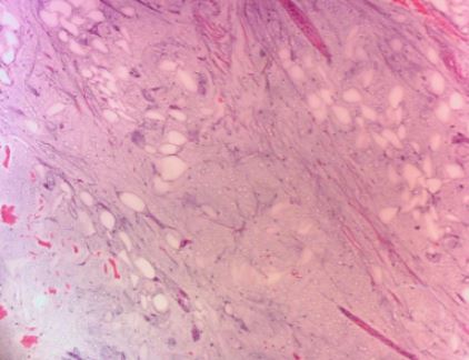
Close interdigitation of adipocytes with myxoid material identifies this as a lipomatous tumor as opposed to a myxoma. Diagnosis – lipoma with myxoid change. Diagnosis confirmed by Sharon Weiss, MD, Emory University.
DISCUSSION
The benign myxoid lipoma should be differentiated from the malignant myxoid liposarcoma histologically. Clinical presentation is a good differentiator. The myxoid lipoma would grow slowly in superficial locations. Liposarcomas present with rapid growth as deep soft tissue lesions. Presence of a capsule around the tumor does not rule out a liposarcoma.

