Posts Tagged ‘dermatopathology’
Treatment Considerations for Melanoma In-Situ
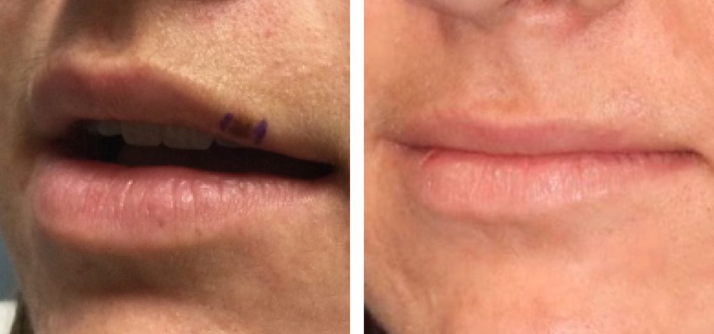
HISTORY A 22-year-old woman presented with left upper lip lesion. Tape testing showed positive for LINC00518 and PRAME. Excisional biopsy was performed showing atypia on the preliminary results. DISCUSSION Management of melanoma in-situ can be especially challenging in cosmetically sensitive areas such as the lip. Traditional management options for melanoma in-situ include surgical excision, topical…
Read More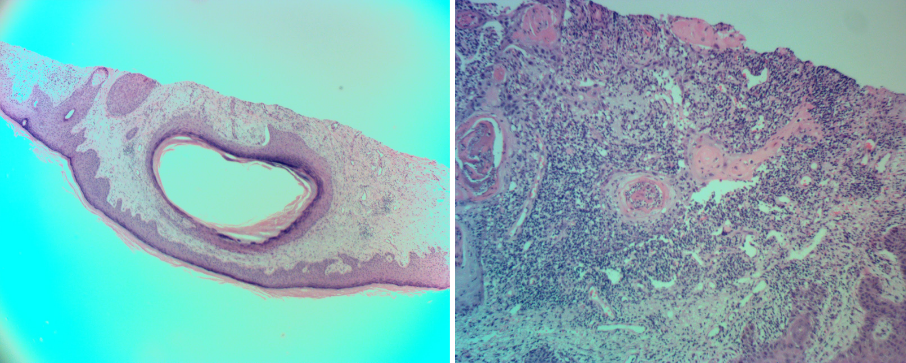
Cystic Squamous Cell Carcinoma of Forearm Skin
HISTORY 74-year-old man presents with 3-month history of painful lesion of the right forearm. Biopsy found cystic squamous cell carcinoma with deep margins involved. Wide local excision was performed achieving clear margins. DISCUSSION Cystic degeneration within squamous cell carcinoma of the skin is not considered an especially malignant finding. These histologic findings may represent changes…
Read More
Management of Recurrent Large Melanoma In Situ
HISTORY 78-year-old man presents with a recurrent melanoma in situ of left cheek in April 2018. Previously, the melanoma in situ was excised in 2001. Patient’s dermatologists performed excision in three stages over a period of 3 weeks to achieve clear margins. Reconstruction of left cheek was performed with a cheek and neck platysma myocutaneous…
Read More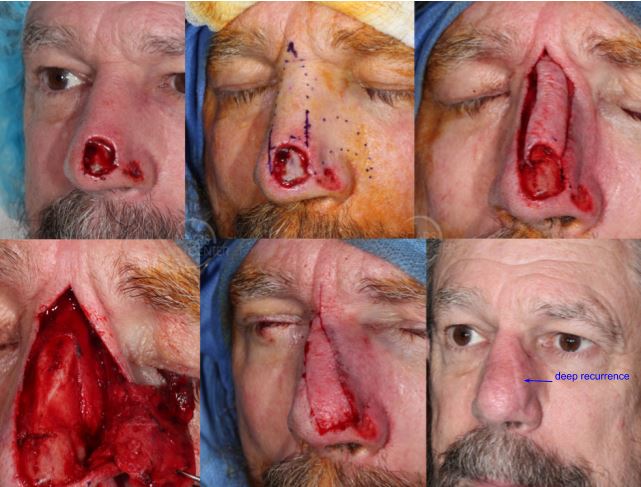
Recurrent SCC of Nasal Tip
HISTORY 71-year-old man presents with recurrence of SCC of nose following Mohs excision on 2-9-17. Partial rhinectomy was performed with incomplete Mohs excision leaving residual positive margins due to patient’s comfort. Two additional excisions of involved margins were done under anesthesia achieving clear margins. First stage reconstruction was performed on 4-11-18. DISCUSSION Management of a…
Read More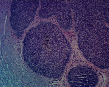
Pigmented BCC of Left Neck
HISTORY 81-year-old man presents with pigmented lesion on left neck. The lesion was excised with 2 mm margins. BCC was found with clear margins. DISCUSSION Pigmented basal cell carcinoma is an uncommon variant of the common skin cancer. Its behavior is that of a non-pigmented basal cell carcinoma. It can be confused with melanoma and…
Read More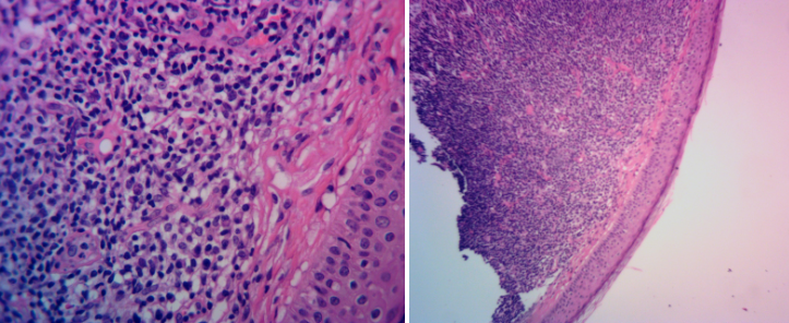
Cutaneous B-Cell Lymphoma of the Ear
HISTORY 49-year-old woman presents with a shiny pink papule of the left ear helix. Histology found atypical lymphoid infiltrate suspicious for a cutaneous B cell lymphoma. The area was treated with Mohs excision and closure. DISCUSSION Primary cutaneous lymphoma is a low-grade and most-common B-cell lymphoma of the skin. These are primary tumors of…
Read More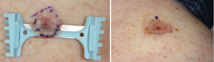
Shave excision of Lower Extremity Carcinomas
HISTORY 67-year-old woman presents with 3 BCCs on right lower extremity diagnosed on shave biopsies. Superficial shave excisions of the carcinomas left close margins within 0.1 mm. Second set of shave excisions found no residual carcinoma. DISCUSSION Lower leg skin carcinomas are often superficial and can be treated with superficial treatments. This patient’s…
Read More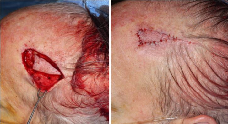
Mitoses in a Dysplastic Nevus
HISTORY 61-year-old man presents with 6-month history of forehead lesion. Biopsy on October 2017 showed atypical nevomelanocytic neoplasm with several mitoses (3/mm2). An excision with 5 mm margins was performed in November 2017 with negative findings. DISCUSSION This melanocytic tumor of uncertain malignant potential can also be called a MELTUMP. These lesions present a treatment…
Read More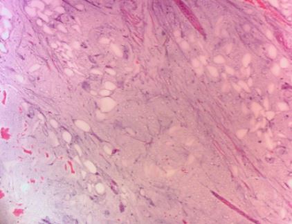
Myxoid Lipoma of Scalp
HISTORY 61-year-old man presents with 40 year history of 2.5 cm mobile lipoma on left scalp vertex. Ultrasound performed on 11-14-17 revealed lipoma. Results from excision of mass showed myxoid lipoma. Consultation was obtained with Emory’s Sharon Weiss, MD, a soft tissue pathology expert. DISCUSSION The benign myxoid lipoma should be differentiated from the malignant…
Read More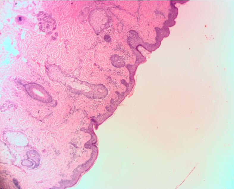
Left Cheek Atypical Spitzoid Nevus
HISTORY 23-year-old woman presents with lifelong history of left cheek mole changing recently. Biopsy in 8/2017 showed compound nevus with severe atypia with spitzoid features. Excision was performed on 2-9-18. The patient a history of atypical mole of the left back removed in 2017. DISCUSSION Spitz nevi are are spindle /…
Read More