Wood’s Lamp Lower Eyelid Melanoma in situ
HISTORY
A 57-year-old woman presents with two year hisory of right under-eye pigmentation. Shave biopsy by an outside office diagnosed MMIS, lentigo maligna type. Evaluated under Wood’s Lamp. Patient will be getting treatment at an outside office.
DISCUSSION
Evaluation of pigmented lesions with blue light
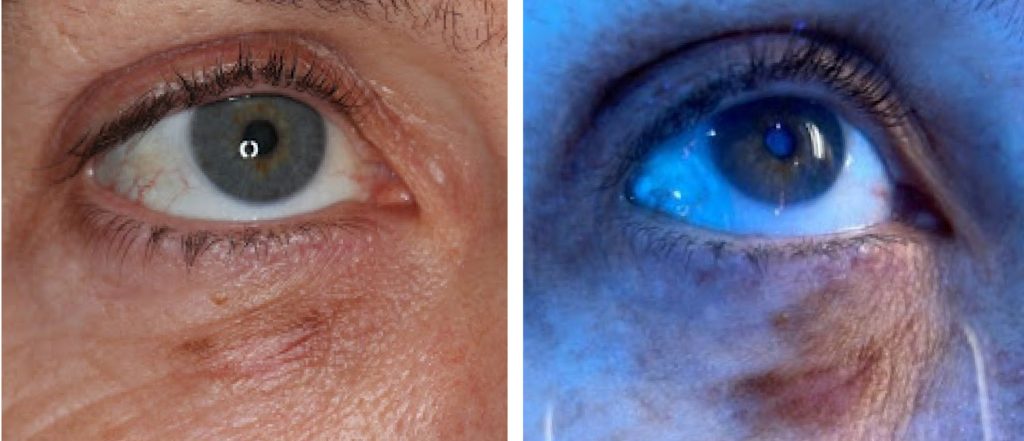
There are multiple aspects to evaluating pigmented lesions, the most important being physical exam. But there is a very helpful tool that providers have in their tool box: a Wood’s Lamp. It is a type of black light emitting a wavelength around 365 nm – the blue edge of the visible light spectrum and includes UV light.
A Wood’s Lamp is a blue fluorescent light that is a very useful tool used frequently in dermatology for the evaluation several diagnoses, such as:
- Pigmented lesions
- Vitiligo
- Tinea Capitis (skin fungal infection)
- Lice
- Pityriasis versicolor
- Erythrasma
- Acne
- Sun damage
Under the Wood’s Lamp, normal skin will look slightly blue, whiter spots are thickened skin, yellow is oily skin, and purple is dehydrated skin. Pigmented lesions will become darker. This helps your clinician identify the extent of the lesion. This tool can be particularly helpful when planning for surgical excision as it allows your clinician to estimate how much tissue will need to removed.
Citations: