Patient Case Studies

Temporoparietal Fasciocutaneous Island Flap for Forehead and Anterior Scalp Reconstruction
ABSTRACT Background: Forehead and anterior scalp large defect reconstruction is challenging and often requires skin grafting. Objective: To measure the advancing distance and the survival of the temporoparietal fascia (TPF) island flap in forehead and anterior scalp reconstruction. Methods: The study design was a retrospective case series. Participants included all patients who had undergone TPF island flap for…
Read More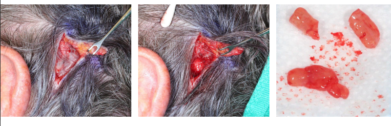
Subcutaneous Intravascular Pyogenic Granuloma of the Scalp of a POEMS Syndrome Patient
ABSTRACT An 82-year-old male with a past medical history of POEMS syndrome presented with a 6-weekhistory of a growing intravascular pyogenic granuloma in the subcutaneous scalp. Biopsyshowed lobular intravascular capillary hemangioma, also known as pyogenic granuloma. Lobularcapillary hemangiomas are most commonly found on the skin and represent some of thecutaneous changes seen within POEMS syndrome.…
Read More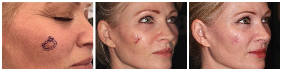
Cerclage Closure Technique
HISTORY A 50 y/o patient presents with a 3 year h/o right cheek growing lesion biopsied by outside office on 11/8/21 showing atypical lentiginous melanocytic proliferation. Incisional biopsy of right malar cheek done on 11/18/21 showing severely atypical lentiginous junctional melanocytic hyperplasia with margins involved. Excision and reconstruction with cerclage of right malar cheek severe…
Read More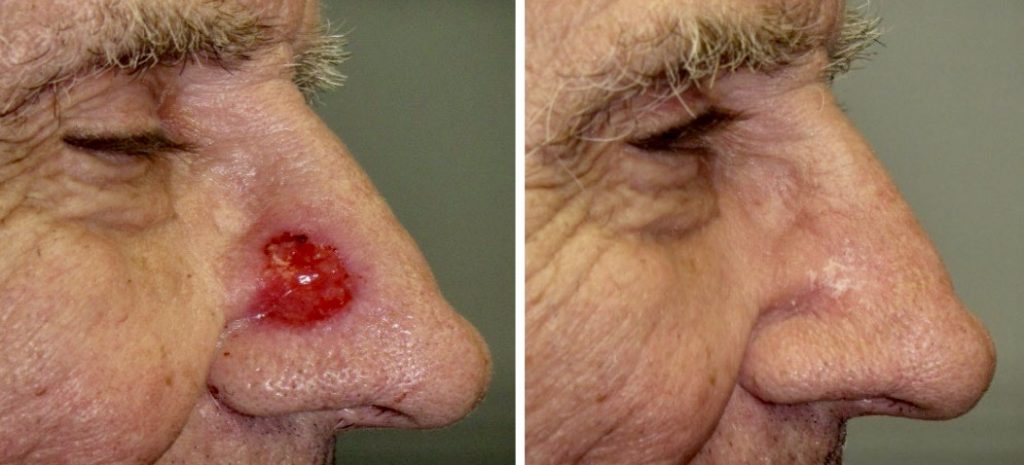
Superficial Radiotherapy: Long Term Follow-Up of Highly Selected Basal and Squamous Cell Carcinomas in Skin Cancer Patients
Superficial radiotherapy (SRT) treatment for non-melanoma skin cancer has been reported to yield variable cure rates. When patients are highly selected, adequate margins of treatment are chosen, and hypofractionation is avoided, cure rates of SRT can approach that of Mohs surgery. SCARS Center’s research article on the subject was recently published in Journal of Dermatology…
Read More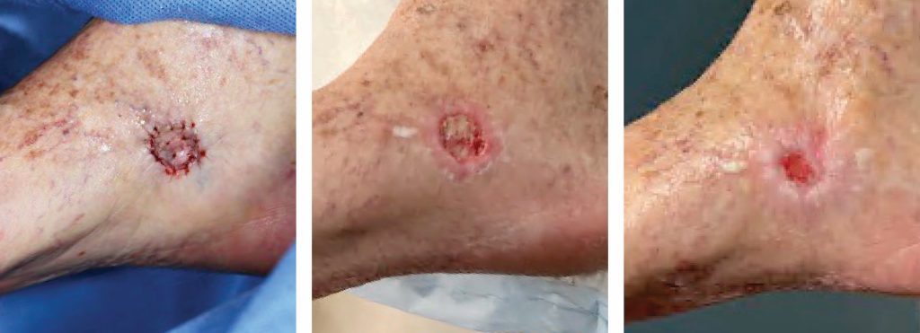
Chronic Open Wound Management with Wound Vac
Chronic open wounds present a lifestyle challenge for patients. In particular, lower leg wounds can take months to fully heal and may require wound care to prevent infection and maintain a healthy, healing wound. A wound vac or “vacuum-assisted closure of wound” can expedite healing. A wound vac decreases the air pressure on the wound…
Read More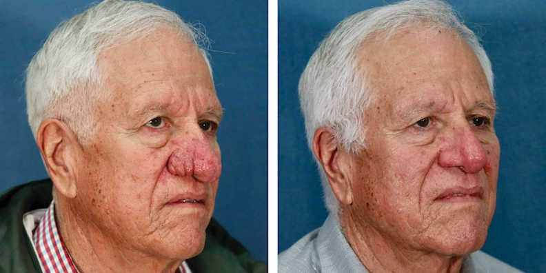
Dermaplaning and Dermabrasion to Treat Rhinophyma
BACKGROUND Rhinophyma is an uncommon form of rosacea that leads to thickening of the nasal skin and enlargement of nasal sebaceous glands. It usually occurs in men over 50. It is slowly progressive and tends to worsen over time, resulting in a bulbous shape of the nose which gradually becomes deforming. Rhinophymatous papules often form,…
Read More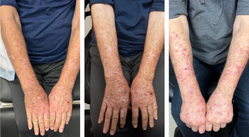
Augmenting Efficacy of Topical 5-FU for Actinic Keratoses and Skin Cancer
Diffuse actinic sun damage can present a challenge for both patients and providers. First line treatment options include topical 5-fluorouracil (5-FU) cream or photodynamic therapy (PDT). Topical 5-FU cream is applied to the affected area for 2-3 weeks creating inflammation, redness, and crusting as the body eliminates the actinic damage. Alternatively, topical levulan is applied…
Read More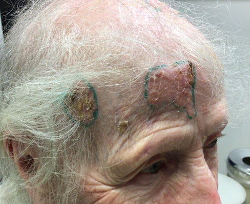
When to do Curettage and Electrodessication (C&D)
HISTORY A 95-year-old woman presents with a 4+ year history of multiple growing lesions of forehead and scalp. These were initially asymptomatic and the patient chose not to seek treatment. She did not present for evaluation and treatment until 2 of the lesions became painful and began to bleed. Shave biopsy of 2 of the…
Read More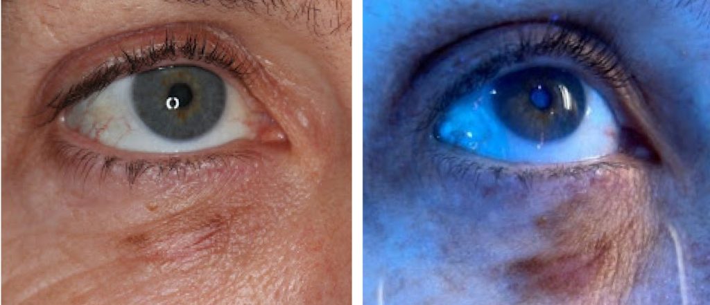
Wood’s Lamp Lower Eyelid Melanoma in situ
HISTORY A 57-year-old woman presents with two year hisory of right under-eye pigmentation. Shave biopsy by an outside office diagnosed MMIS, lentigo maligna type. Evaluated under Wood’s Lamp. Patient will be getting treatment at an outside office. DISCUSSION Evaluation of pigmented lesions with blue light There are multiple aspects to evaluating pigmented lesions, the most…
Read More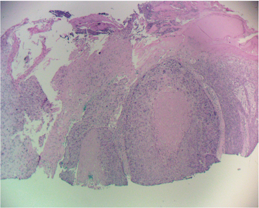
Management of Invasive Squamous Cell Carcinoma Arising in Bowen’s Disease
A patient presented to the SCARS Center with a nine month history of a biopsy proven Bowen’s disease of the left second toe. Mohs surgery discovered areas of invasive squamous cell carcinoma arising from Bowen’s disease within the lesion. After completing Mohs surgery, additional deep soft tissue and phalanx bone were resected to insure tumor…
Read More