Posts Tagged ‘melanoma in situ’
Treatment Considerations for Melanoma In-Situ
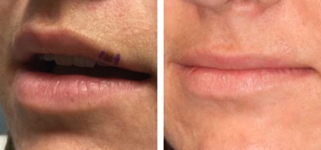
HISTORY A 22-year-old woman presented with left upper lip lesion. Tape testing showed positive for LINC00518 and PRAME. Excisional biopsy was performed showing atypia on the preliminary results. DISCUSSION Management of melanoma in-situ can be especially challenging in cosmetically sensitive areas such as the lip. Traditional management options for melanoma in-situ include surgical excision, topical…
Read MoreManaging Lentigo Maligna Melanoma
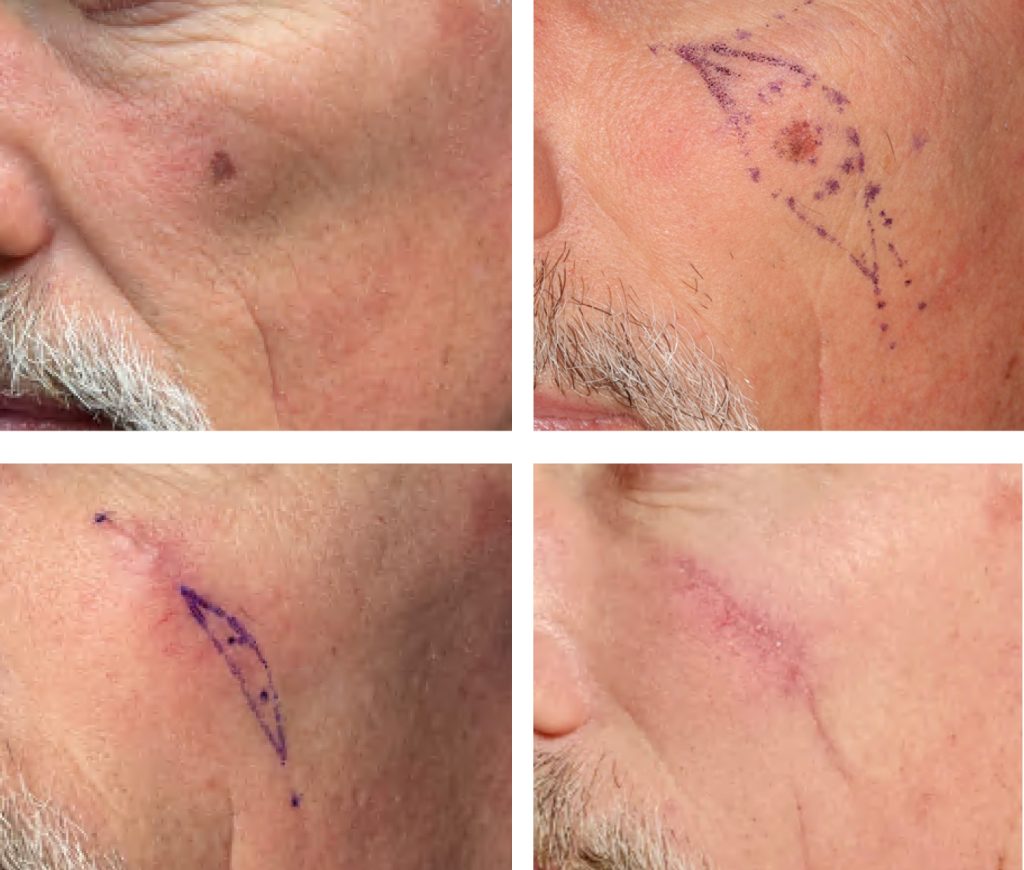
HISTORY A 56-year-old man presented with complaint of lifelong history of darkening, growing left lesion of the left cheek. Biopsy on 09/24/21 showed melanoma in-situ. Excision with 5-6mm margins and closure of left cheek melanoma performed on 12/2/21 showed positive margins at 3-4 o’clock margins. Woods lamp evaluation and re-excision performed on 1/7/22. DISCUSSION Lentigo…
Read MoreAtypical Nevus – a Melanoma Precursor? Not Exactly
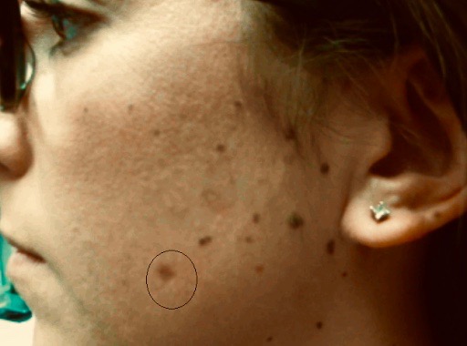
HISTORY A 27-year-old woman presented with many-year history of left cheek nevus. The lesion grew in size and changed in color. Shave biopsy showed a nevus with moderate atypical melanocytic hyperplasia. Residual pigmentation of the nevus and involved histologic margins remained after the biopsy. DISCUSSION Are all atypical melanocytic lesions pre-melanomas? Should dysplastic lesions be…
Read MoreManagement of Recurrent Large Melanoma In Situ
HISTORY 78-year-old man presents with a recurrent melanoma in situ of left cheek in April 2018. Previously, the melanoma in situ was excised in 2001. Patient’s dermatologists performed excision in three stages over a period of 3 weeks to achieve clear margins. Reconstruction of left cheek was performed with a cheek and neck platysma myocutaneous…
Read MoreMapping of Recurrent Melanoma In Situ of Leg
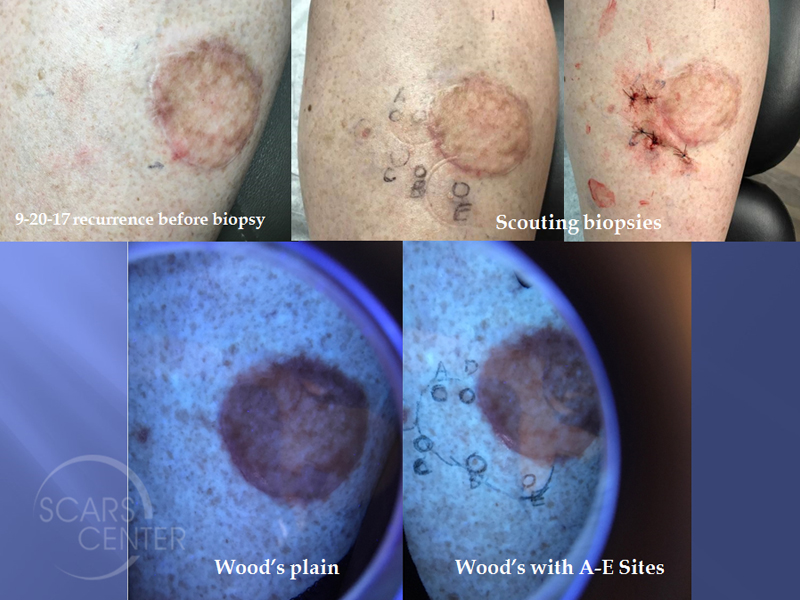
HISTORY 50-year-old woman presents with recurrent melanoma in situ of left leg. First biopsied on 9-2-16, the lesion was excised on 10-14-16 and showed extensive residual melanoma in-situ with radial margins involved from 12- 3 o’clock. The defect was reconstructed at the time with a full thickness skin graft. Second excision cleared the involved…
Read MoreRight Ear Melanoma In Situ with Depth Uncertainty – The role of Histopathologic Discordance in Clinical Decision Making
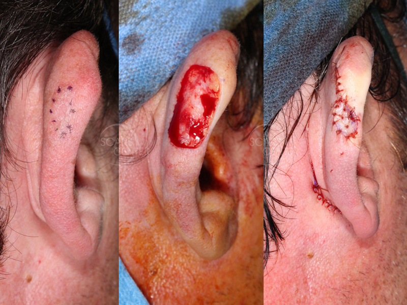
HISTORY 40-year-old man presents with a 2 month history of right posterior ear pigmented papule. Shave biopsy on 9-18-17 showed malignant melanoma at least in situ. Excision with 5 mm margins on 10-9-17 confirmed residual melanoma at least in situ with clear margins. DISCUSSION Lack of superficial invasion cannot be definitely ruled out after a…
Read MoreChasing Margins of Melanoma In Situ of Back
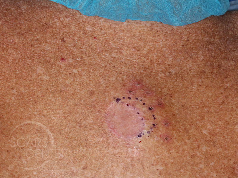
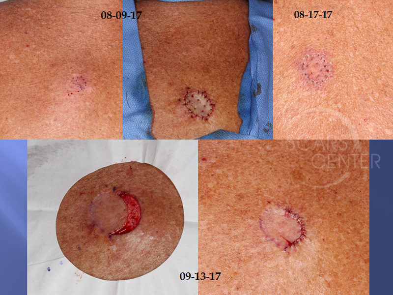
HISTORY 69-year-old man presents with melanoma in situ of the upper back. A biopsy on 6/15/2017 showed malignant melanoma at least in-situ. Excision with 5 mm margin and skin graft closure on 8/9/2017 found residual melanoma in situ with at 2-4 o’clock. Additional 4mm margin excision on 9-13-17 still found residual lesion at 1-3…
Read MoreRECURRENT MELANOMA
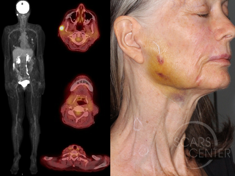
HISTORY 70-year-old woman presents with metastatic melanoma in the right parotid gland region of 1 year duration. Diagnosis was made with a core needle biopsy on 09-08-17. Patient also has small palpable right cervical lymph nodes with SUV 1.6 and 2.0. Patient history of melanoma began in 2001 as a pink macule of the…
Read MoreCHEEK AND EYELID RECONSTRUCTION WITH MULTIPLE PARTIAL ISLAND FLAPS
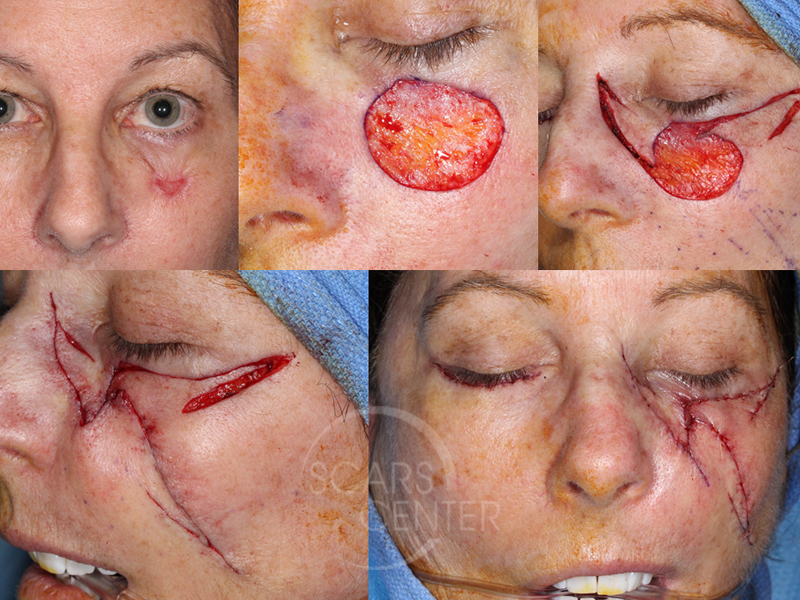
HISTORY 61-year-old woman presents with several year history of pigmented left cheek and lower lid lesion. She was initially treated with liquid nitrogen 15 months ago. A biopsy was performed 6-8-17 and revealed melanoma in situ. Excision with close margins performed 6-16-17. Clear margins found on LPMG reading. Second opinion from UCSD was suspicious for…
Read MoreADDITIONAL MARGIN EXCISION AFTER SKIN GRAFTING
DISCUSSION This series of photographs represents an approach to margin management of melanoma in situ. Some melanoma in situ margins are difficult to assess clinically, as is the case in our first series with extensively pigmented skin. In some settings, taking 7-9 mm margins exposes the patient to significant healing issues, as the second case…
Read More
